Ultrasound 3D Tomography
Hello We are team Oculus.
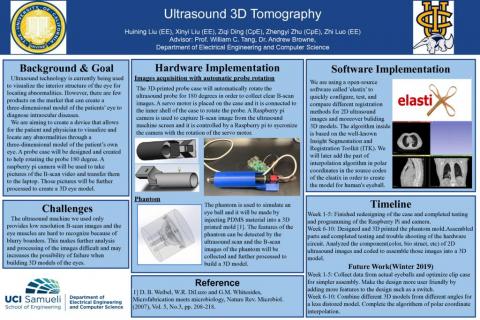
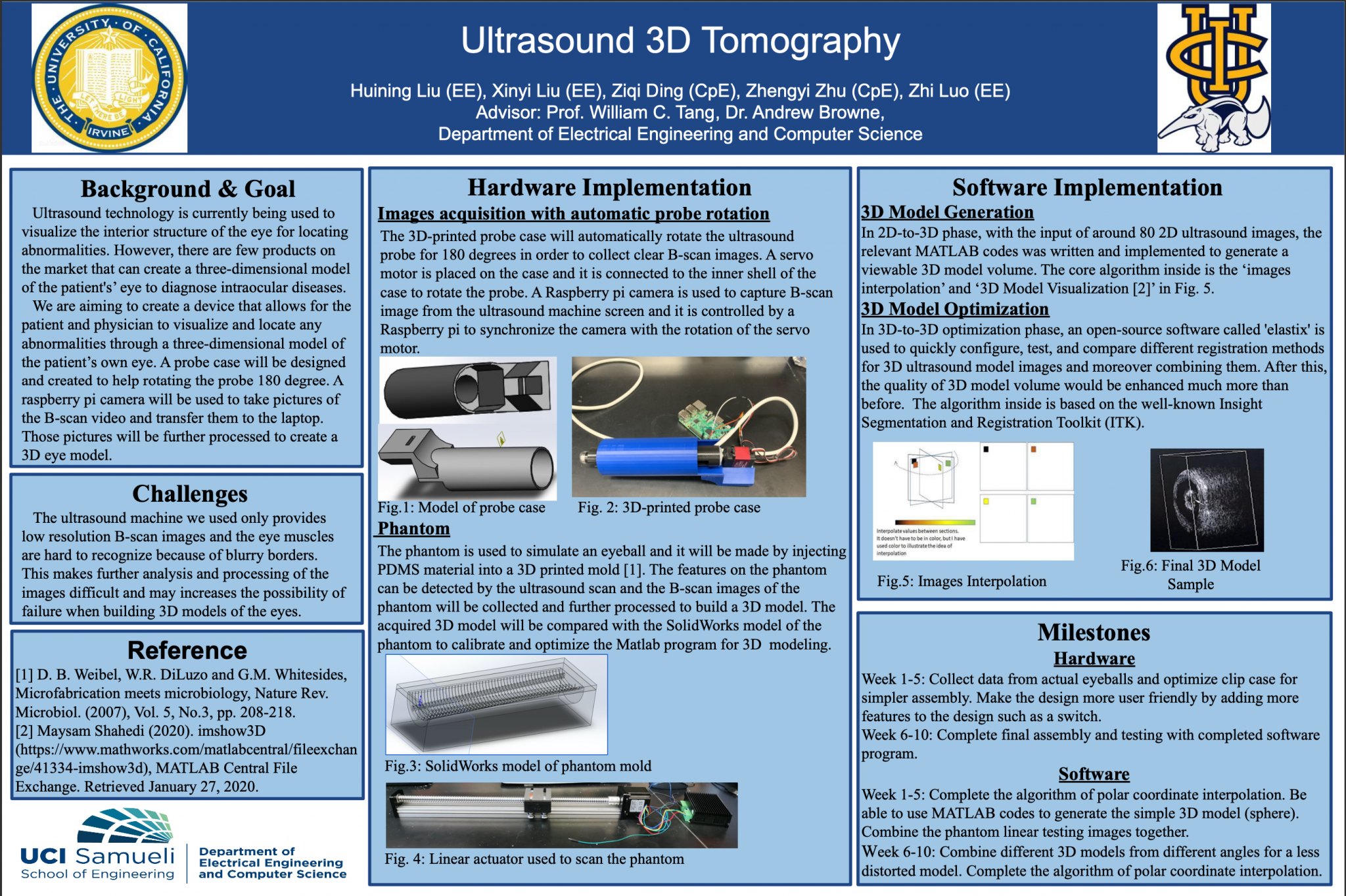
Ultrasound technology is currently being used to visualize the interior structure of the eye for locating abnormalities. However, the major limitation of this approach is that most of the ocular ultrasonography viewing devices can only display two-dimensional scans that provides the physician with limited spatial information, complicating the process of diagnosing the patient. And there are few products on the market that can create a three-dimensional model of the patients’ eyes to diagnose intraocular diseases. There have been documented attempts to develop systems that can produce 3-D models and are suitable for use on the human body. However, various obstacles have slowed down the progression and success of these systems. Since the sequence of the acquired tomographic images is crucial to the formation of the desired 3-D model, the geometry of the body part must be known in order to avoid any distortions, and the images must be acquired quickly to minimize any patient motion. Additionally, the existing B-scan technology does not provide high resolution scan images. Therefore, the acquired 3-D models are often inaccurate and blurred. As a result, these systems were generally unsuccessful due to either the 3-D imaging devices being too slow or because complicated instrumentation is required.
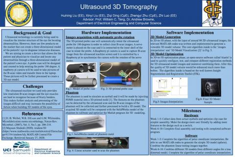
We are aiming to create a device that allows for the patient and physician to visualize and locate any abnormalities through a three-dimensional model of the patient’s own eye. The end of the goal is to create a device including a holder for the ultrasound machine probe and it will be able to automatically rotate the probe 180 degrees forward and back according to the physicians’ scanning procedure. There will also be a camera that will take photos of the B-scan video from the ultrasound machine, while transferring the photos to computer. The B-scan photos will further be processed and integrated into a 3-D model to assist the physicians annotating anatomical locations and lesions in the eye. A phantom of an eye will be used for 2-D image data collection. The phantom will be made by injecting PDMS material into a 3-D printed mold. The acquired 3-D model will be compared with the phantom to identify any distortion and inaccuracy.
This project was the senior design project of a group of biomedical engineering students at the University of California, Irvine from 2018 Fall quarter to 2019 Spring quarter. In this quarter, we took over their work and continued the unfinished work and optimized the existing prototype. The previous group have designed a case to hold the ultrasound probe and used a stepper motor to rotate the probe. However, the torque provided by the stepper motor was not large enough so the rotation was unsuccessful. In this quarter, we have substituted the stepper motor with a servo motor that is more powerful and more accurate so that the servo motor can smoothly rotate the probe with small steps to capture B-scan images. Also, we used Raspberry Pi to synchronize the rotation of the probe and the photo taken by the Raspberry Pi Camera. A crescent shaped phantom have been made to simulate the back of the eye. A set of 2-D B-scan images of the phantom have been collected using the existing images for further image processing.